
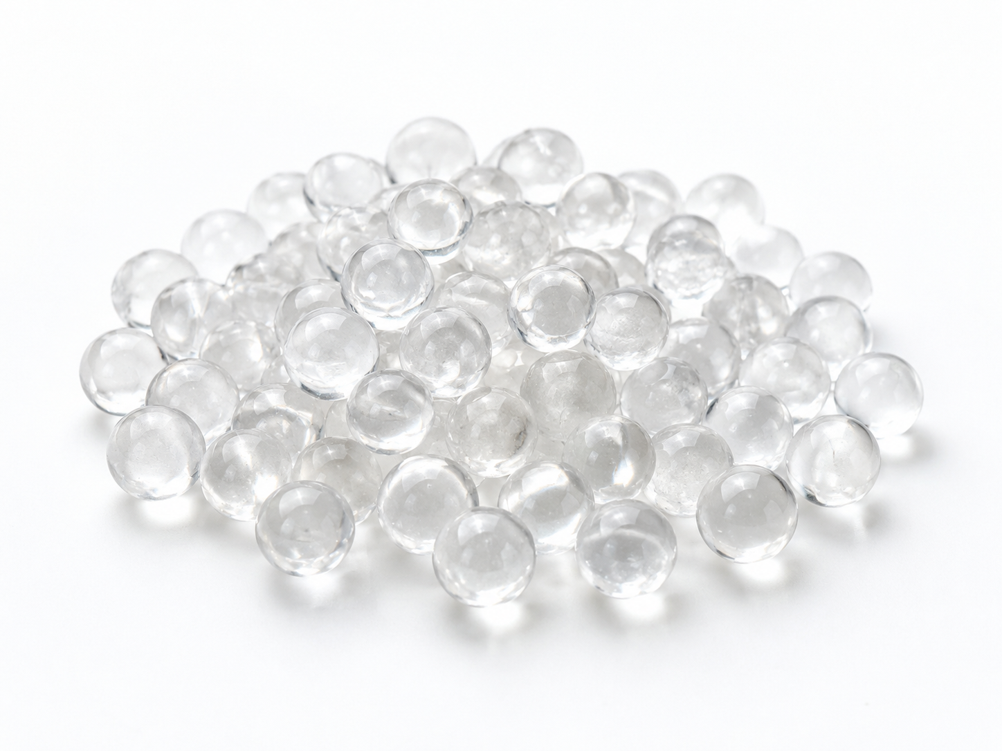
Cross-sectioning is the most direct way to see inside a siliphos ball or polyphosphate glass sphere: you cut or grind through the ball, polish the exposed face, and examine it under a microscope. Done correctly, a cross-section reveals internal voids, cracks, unmelted raw material, core-to-rim composition differences, and other defects that surface inspection, weight checks, and visual sorting cannot detect. Done carelessly, it creates artifacts that look like defects but are actually damage from cutting, heat, or water. This article explains how to prepare and read a cross-section so you can judge whether an incoming batch of polyphosphate media is internally sound, and how to connect what you see to real performance in a water-treatment device.
What a Cross-Section Can and Cannot Tell You
A cross-section answers structural questions: is the interior solid or porous? Are there cracks? Is the material uniform from center to surface? Are there embedded particles or unmelted batch?
It does not answer every question. A cross-section will not tell you the full chemical formulation, the dissolution rate, the scale-inhibition dose response, or whether the material is acceptable for drinking water. Those require analytical chemistry, dissolution testing, and market-specific compliance review of the exact product. Treat the cross-section as one instrument in a set, not a complete quality verdict.
It is also worth separating functions before you begin. Polyphosphate media of this family are generally used for scale inhibition — they release phosphate species that interfere with carbonate scale formation. They are not water softeners, not filters, and not disinfectants. A crack or void matters because it changes how the ball dissolves and whether it sheds particulate matter, not because it affects softening capacity.
Why Internal Defects Matter in Service
Glassy polyphosphate balls are usually produced by melting a blend of phosphate salts and quenching the melt into spheres. That process can trap gas, produce thermal stress, or leave localized regions richer in one component than another. In a filter housing or dosing cartridge, the consequences depend on the defect:
- Connected porosity or open cracks increase the exposed surface area, which can accelerate dissolution and shorten the useful life of the bed in an uncontrolled way.
- Large internal voids weaken the ball mechanically; under flow, vibration, or the weight of a packed bed, it may fracture and generate fines.
- Unmelted or crystalline inclusions may dissolve at a very different rate from the surrounding glass, leaving behind a skeleton of slowly soluble material and causing uneven release.
- Core-to-rim gradients can mean the ball does not dissolve uniformly, which complicates any attempt to predict dosing from bed weight alone.
None of these automatically makes a batch unusable. The question is whether the defect population is consistent with the dissolution profile your equipment was designed around. Cross-sections let you see whether a supplier’s process is under control batch to batch.
Preparing a Cross-Section That You Can Trust
1. Sample with a plan, not a handful
Take a stratified sample rather than scooping from the top of a drum. Draw balls from the top, middle, and bottom of the container, and from more than one container if the shipment is large. Record the batch or lot identity for each specimen. If you only section one or two balls, you are describing those balls, not the batch.
2. Section through the center
A cut that misses the geometric center makes internal voids appear smaller than they are and can hide a core structure entirely. For critical evaluation, prepare at least two planes — ideally two perpendicular central planes — so you can see whether features are radially symmetric (often a sign of the quenching process) or randomly distributed (often a sign of melt contamination or gas entrapment).
3. Control heat, water, and mechanical damage
Polyphosphate glass is sensitive to both heat and moisture, so sectioning method matters:
- Use a low-speed precision saw with a fine abrasive blade and light feed pressure to limit heat generation.
- Avoid water-based coolants if possible. Water can dissolve or hydrate the freshly exposed surface and round off the edges of pores, producing a false “smooth” appearance. A non-aqueous lubricant or a short, controlled dry cut is generally safer.
- If a crack is already present, vacuum-impregnate the specimen with a low-viscosity epoxy before cutting so the fracture does not crumble or smear during polishing.
- Polish progressively on fine abrasives. Soft glass phases can smear and fill small voids, so inspect after each step and clean the surface in a way that does not dissolve the sample.
4. Choose the right imaging method
| Method | Best for | Limitations |
|---|---|---|
| Stereo microscope (reflected light) | Voids, cracks, color banding, gross inclusions at low magnification | Limited resolution; depth of field can mislead on pore size |
| Reflected-light metallography | Polished-surface detail, void area measurement, crack path | Requires careful preparation; smearing artifacts are common |
| Polarized light | Detecting crystalline inclusions in an otherwise amorphous glass | Does not identify the crystal phase by itself |
| SEM with EDS | Elemental mapping across core-to-rim gradients, inclusion chemistry | Semi-quantitative without standards; vacuum and coating requirements |
| Micro-CT (non-destructive) | Three-dimensional void and crack mapping before cutting | Resolution limits for fine cracks; weak contrast between similar-density phases |
Defect Types and What They Usually Indicate
| Feature seen in section | Typical appearance | Likely origin | Practical concern |
|---|---|---|---|
| Spherical voids | Round, dark, smooth-walled pores | Gas entrained during melting or quenching | Reduced strength; possible increase in surface area if interconnected |
| Cracks | Sharp linear features, often radial | Thermal shock during quenching; mechanical handling | Fines generation, unpredictable dissolution |
| Crystalline inclusions | Bright, birefringent particles under polarized light | Incomplete melting or devitrification | Uneven dissolution; residual insoluble residue |
| Core/rim contrast | Color or texture difference between center and edge | Incomplete homogenization or differential cooling | Non-uniform release rate; needs confirmation by EDS |
| Opacity bands | Concentric cloudy zones | Phase separation or hydration during storage | Possible surface degradation; check storage conditions |
These interpretations are indicative, not diagnostic. A bright particle under polarized light could also be an artifact from polishing debris, and a dark spot could be a pulled-out inclusion rather than a void. Confirm every observation with a second technique before writing it into a rejection report.
From Images to Decisions
Qualitative inspection is useful for screening, but purchasing and quality decisions benefit from numbers. If you have image-analysis software, measure:
- Void area fraction — total void area divided by the section area.
- Maximum void diameter — a single large void is often more consequential than many small ones.
- Crack length per unit area and whether cracks reach the surface.
- Inclusion count and size distribution per unit area.
- Radial uniformity — element intensity or optical density plotted from center to rim.
Do not invent acceptance limits from these numbers alone. Any threshold should be agreed with the supplier and tied to a functional test, because the relationship between void fraction and dissolution behaviour depends on the formulation and the application conditions. The most defensible approach is to cross-section a batch you already know performs acceptably, establish a baseline, and compare new lots against that baseline using the same preparation method.
Limits, Artifacts, and Common Mistakes
- Water-based cutting and mounting. This is the single most common source of false results with soluble phosphate glass. Use non-aqueous methods or keep water contact minimal and documented.
- Off-center sections. They understate void size and can make a cored structure invisible.
- Sample size of one. Batch variability cannot be judged from a single ball.
- Confusing appearance with performance. A cross-section does not measure dissolution rate. Pair it with a dissolution or conductivity test under controlled conditions.
- Comparing unlike materials. “Siliphos,” “siliphos crystal,” “phosphate crystal,” and “polyphosphate media” are used loosely in the market and do not always describe the same composition or manufacturing route. Confirm what you are actually comparing.
- Ignoring safety. Cutting and grinding generate fine phosphate-containing dust. Use appropriate ventilation and personal protective equipment, and follow your site’s handling rules.
Non-Destructive Alternatives and When to Use Them
Micro-CT can map internal voids and cracks in three dimensions without destroying the sample, which makes it valuable for expensive or archival specimens and for tracking the same ball through a dissolution trial. Its weakness is resolution: hairline cracks and low-contrast phase boundaries may not appear. A practical workflow is micro-CT first, then targeted cross-sectioning of the specimens that show the most interesting features, so destructive work is spent where it will be most informative.
For drinking-water projects, remember that structural quality and regulatory suitability are separate questions. A clean cross-section says nothing about whether a specific product may be used in potable water in a given market. That determination depends on the exact product, the application, and local requirements, and must be verified with the supplier and the relevant authority.
Frequently Asked Questions
Do voids in a polyphosphate ball always cause problems?
No. Small, isolated, closed voids often have little effect on dissolution or strength. Risk rises when voids are large, connected to the surface, or clustered in one region, because they change the effective surface area and mechanical integrity.
Can I evaluate internal quality without cutting the balls?
Partly. Micro-CT and, in some cases, ultrasound can detect gross internal features non-destructively, but fine cracks and compositional gradients are usually better resolved on a polished cross-section.
How many balls should I section per batch?
There is no universal number. It depends on batch size, the variability you observe, and the sampling plan you agree with your supplier. Start with stratified samples across the container and increase the count if you see inconsistent results between specimens.
Does a uniform cross-section prove the material is suitable for drinking water?
No. Internal uniformity is a manufacturing quality indicator. Drinking-water suitability must be established separately for the exact product and application in the destination market.
Is a color difference between the core and the rim a defect?
Not necessarily. Differential cooling can produce optical differences without a meaningful compositional change. Confirm with elemental mapping before treating it as a nonconformity.
Specifying and Buying with Cross-Section Data in Mind
If internal quality matters for your application, put it in the specification rather than discovering it after delivery. Useful items to agree with a supplier include the sampling plan, the sectioning and mounting method (so results are comparable between parties), the imaging methods to be used, the defect features to be reported, and the functional test that will be used alongside the structural inspection. Ask whether routine process controls — melting, homogenization, quenching — are documented, and whether inspection records are available for the lot you are purchasing.
ENVO CHEMICAL can discuss cross-section inspection criteria and material selection for siliphos-type and polyphosphate media when you share the application details. To get a useful answer rather than a general one, send your intended use (scale inhibition, dosing cartridge, or other), source-water conditions such as hardness and temperature range, equipment type and flow rate, required ball size or specification, expected quantity, destination market, and the specific technical objective of your inspection. With those details, we can respond to the actual problem instead of a generic product list.
