
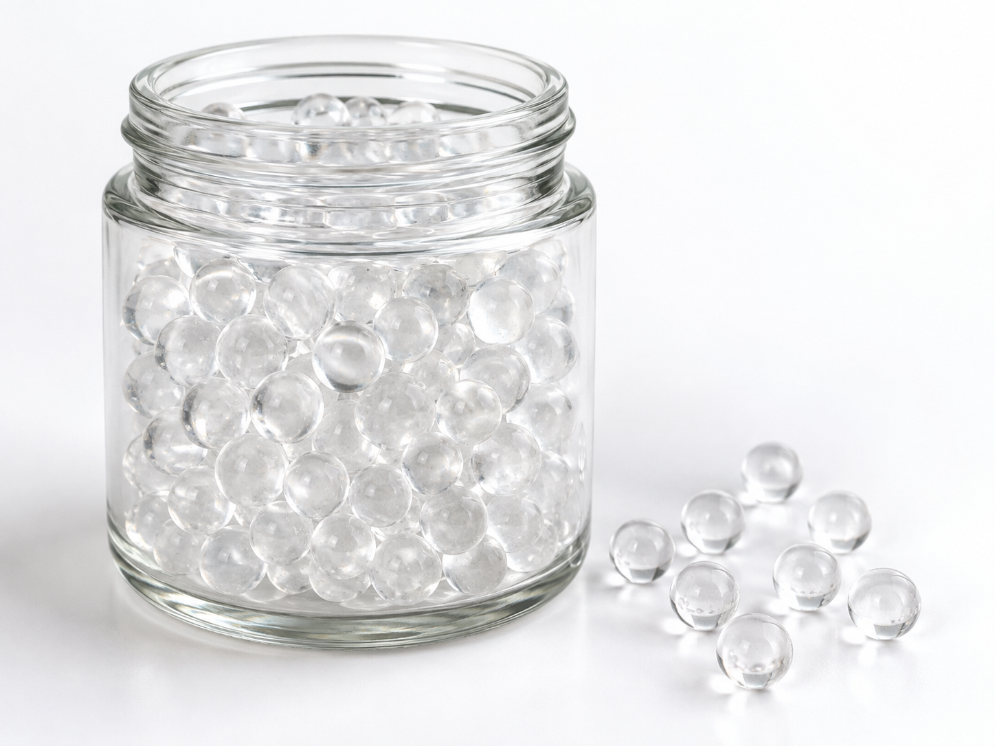
Microscopy is the quickest way to find out whether the inclusions inside a batch of polyphosphate glass balls are ordinary melt bubbles or something that will change how the product dissolves. A representative sample is mounted, examined under reflected and transmitted light, and each inclusion is classified by shape, optical behaviour, and—when needed—elemental composition. The method is fast and inexpensive, but only if sample preparation respects one important fact: polyphosphate glass dissolves in water, so conventional water-based polishing and cleaning will destroy the very features you are trying to measure.
This article is written for the people who actually have to make a decision with that information: quality engineers at water-treatment equipment manufacturers, incoming-inspection staff at importers and distributors, and process engineers evaluating whether a supplier’s glass balls will behave consistently in a dosing or scale-control installation.
What counts as an inclusion in a polyphosphate glass ball
An inclusion is any feature inside the glass that is not part of the continuous glass network. In slow-dissolving polyphosphate balls, the common categories are:
- Entrained gas bubbles. Usually spherical, sometimes elongated or clustered, formed when gas is trapped in the melt. These are the most common feature and are frequently benign.
- Crystalline phases. Devitrification products, unmelted batch material, or crystalline phosphate phases that form on cooling. These may have a very different solubility from the surrounding glass and can change the way a ball breaks down.
- Refractory or foreign particles. Sand, alumina, zirconia, or other furnace and handling debris that never melted into the glass.
- Metallic particles. Fragments from moulds, crushers, screws, or conveying equipment. These are usually distinct under reflected light and easy to confirm with elemental analysis.
- Compositional heterogeneities. Streaks, droplets, or phase-separated regions that are still glassy but not chemically uniform. These do not show birefringence and are the hardest to detect without chemical mapping.
For clarity of terminology: Siliphos is a brand name associated with slow-dissolving polyphosphate glass spheres, and “siliphos crystal” is often used loosely in the trade for this whole product family. Not every polyphosphate glass ball is chemically or physically identical, so inclusion findings from one supplier’s product should never be assumed to apply to another’s.
Why inclusions matter to the end user, not just the glassmaker
Polyphosphate glass balls work by slowly releasing phosphate species into the water stream. That release is controlled by the surface area that is exposed as the ball dissolves. If inclusions change how the ball breaks apart—by creating preferential dissolution paths, by leaving a residue, or by causing the ball to crumble instead of shrinking evenly—then the phosphate feed rate becomes less predictable.
Practical consequences reported by equipment manufacturers and maintenance teams include:
- Residue and particulate carry-over. Insoluble refractory or metallic particles can pass downstream and contribute to turbidity, or settle in low-flow zones.
- Blocked injectors, valves, and screens. Fine fragments from uneven dissolution can accumulate in narrow passages.
- Inconsistent dosing. If a proportion of the bed dissolves faster or slower than expected, the delivered phosphate concentration drifts.
- Bypass or channelling. Balls that soften, cake, or collapse unevenly allow water to take a preferred path through the bed instead of contacting the full volume.
It is worth being precise about function here, because the terminology is often blurred. Polyphosphate glass dosing is primarily a scale-inhibition and sequestration measure: it interferes with the formation and deposition of certain mineral scales and can hold small amounts of hardness ions in a dispersed form. It is not water softening in the ion-exchange sense, it does not remove hardness from the water, it is not a filtration process, and it is not disinfection. If the actual problem is high hardness, iron, or biological growth, microscopy results on glass balls will not address it.
Which microscopy technique answers which question
| Technique | What it reveals | Sample form | Main limitation |
|---|---|---|---|
| Stereomicroscopy (reflected light) | Ball shape, surface condition, colour variation, visible bubbles and opaque specks after fracturing | Whole balls, fractured pieces | Limited resolution; interior only visible after breaking or sectioning |
| Polarised light microscopy (PLM), transmitted | Crystalline versus glassy inclusions; birefringence; strain patterns; particle shape and habit | Crushed grains or thin sections in immersion medium | Strain birefringence in glass can be mistaken for crystallinity without experience |
| Immersion / refractive index oils | Relative refractive index of a particle, supporting identification of the phase | Grain mounts | Requires a reference oil set and careful technique; not a chemical identification on its own |
| SEM-EDS | Elemental composition of individual inclusions and their surrounding glass | Polished or fractured mounts, coated | Phosphate glasses can be beam-sensitive; results are semi-quantitative without standards |
| X-ray diffraction (XRD) | Confirmation of which crystalline phases are present, when enough material can be separated | Powder or hand-picked inclusions | Bulk method; trace phases may be missed; not a substitute for imaging |
| Hot-stage microscopy | Softening behaviour and how inclusions respond as the glass dissolves in a controlled liquid | Small fragments under a coverslip | Small-scale behaviour does not automatically predict full-bed performance |
In most incoming-inspection programmes, stereomicroscopy plus PLM answers the routine questions, and SEM-EDS is reserved for inclusions that look unusual or appear in a location where they could reach the water stream.
Sample preparation is where most investigations succeed or fail
Polyphosphate glasses are relatively soft and water-soluble. That single material property dictates the preparation rules.
- Keep water out of the process. Use isopropanol, anhydrous ethanol, or a non-aqueous immersion medium for cleaning, mounting, and polishing. Even brief water contact can round edges, dissolve fine crystalline material, and create surface artefacts that look like inclusions.
- Sample representatively. Use a riffle splitter, cone-and-quartering, or a documented random selection across the batch, not the handful that happens to be on top of the drum. Inclusion content varies with position in a melt and with particle-size fraction.
- Record the size fraction. Crush and sieve into defined fractions. A high inclusion count in the fines may say more about crushing than about the original product.
- Control the crushing energy. Aggressive crushing produces conchoidal fracture surfaces, fine debris, and metal contamination from the tool. Use a clean agate or ceramic mortar, or a mill that does not shed iron or carbide.
- Mount in a medium close to the glass refractive index if you need to see internal features clearly. A close match reduces surface scatter and lets you look into the grain rather than at it.
- Polish lightly, with non-aqueous lubricant, and clean the sample immediately before examination. Soft phosphate glass smears easily, and a smeared layer can hide or imitate inclusions.
- Document magnification, illumination mode, and location for every image. Inclusion work is comparative; without consistent conditions, batch-to-batch comparison is meaningless.
A practical workflow for a batch investigation
- Define the question first — for example, “why did this batch leave more residue than the previous one?” — because that determines the sampling plan.
- Examine whole balls under a stereomicroscope at low magnification and note colour, transparency, surface crazing, and any visible internal features.
- Fracture a subset and examine the fresh interior under reflected light to count bubbles and locate opaque particles.
- Crush a representative portion and prepare immersion mounts in a non-aqueous medium.
- Examine under transmitted plane-polarised and cross-polarised light. Classify each inclusion as gas, crystalline, amorphous heterogeneity, or opaque foreign particle.
- Select anomalous particles for SEM-EDS, and if a crystalline phase is present in useful quantity, confirm it by XRD.
- Score the batch: inclusion types, size range, approximate count per unit mass, and whether inclusions are sealed inside the glass or exposed at the surface.
- Correlate the findings with a controlled dissolution test and with field performance data from the same batch.
Step eight is the one that turns microscopy from a curiosity into an engineering tool. Inclusion data alone does not predict dissolution rate; it becomes useful when linked to measured dissolution behaviour and to what the customer actually observed in the field.
Reading the results: normal variation versus a genuine concern
Not every inclusion is a defect. Small, well-rounded, isolated gas bubbles are a normal feature of melted and cast glass and are often acceptable. The findings that usually deserve follow-up are:
- Large or clustered bubbles concentrated in one region of a ball, which may indicate a filling or cooling problem.
- Crystalline inclusions with sharp outlines, especially in high numbers, which can point to devitrification or incomplete melting.
- Opaque angular particles that do not dissolve in the immersion medium — possible refractory or sand contamination.
- Metallic specks, which usually indicate wear from processing equipment and should trigger a review of the crushing and conveying line.
- Inclusions exposed at the surface, which have a more direct path into the water stream than those fully encapsulated in glass.
A useful rule is to compare against a retained reference sample from an accepted batch, examined under identical conditions. Absolute thresholds are less meaningful than a documented baseline plus a defined action level.
What microscopy cannot tell you
Microscopy and its complementary techniques describe structure and, with EDS, local elemental composition. They do not establish:
- The dissolution rate or expected service life of the product under your specific water conditions.
- Whether the material is suitable for potable water. That depends on the exact product, the application, and the regulatory requirements of the destination market, and must be confirmed through the appropriate migration and certification documentation — not from appearance or from a “food grade” description alone.
- Performance limits with respect to temperature, pH, hardness, flow rate, or dosage. Those must come from the supplier’s verified data and from your own application testing.
- The cause of a scaling or corrosion problem elsewhere in the system. Microscopy of glass balls is one line of evidence, not a system diagnosis.
Frequently asked questions
Are bubbles inside polyphosphate glass balls a defect?
Usually not. Small, isolated, rounded bubbles are a normal consequence of glass melting and handling. They become a concern when they are large, clustered, or concentrated in a way that suggests the ball may break down unevenly.
Can I identify an inclusion without SEM-EDS?
Often yes. Shape, colour, relief, and behaviour under cross-polarised light distinguish gas bubbles, crystalline phases, and opaque foreign particles in many cases. EDS is what confirms the elemental identity of an unfamiliar particle, so it is normally used for the few inclusions that matter most.
Why must sample preparation avoid water?
Because the product is designed to dissolve in water. Water-based cutting, grinding, or rinsing will alter the surface, dissolve fine crystalline material, and introduce artefacts that can be mistaken for inclusions or that hide real ones.
Does a clean microscopy result guarantee good performance?
No. A batch can be free of visible inclusions and still behave differently because of glass composition, thermal history, or particle size distribution. Microscopy is a structural check; it should be combined with dissolution testing and, where relevant, with field performance records.
Specifying what you need from a supplier
If you are writing an incoming-inspection specification or asking a supplier to investigate a complaint, the following items are worth requesting in writing: the size fraction supplied, the intended application and equipment type, any agreed dissolution or residue test method, the reference sample used for comparison, and the identity of any certification required for the destination market. Keep microscopy findings and chemical or regulatory documentation separate — they answer different questions.
ENVO CHEMICAL supplies polyphosphate glass balls and related water-treatment materials, and can discuss how inclusion and dissolution behaviour is characterised for a specific application. To move quickly, send the intended application, source-water conditions (hardness, pH, temperature, and any iron or manganese), equipment type and flow rate, the specification or size fraction you require, estimated quantity, destination market, and the technical objective you are trying to meet. With those details, it is possible to confirm what is relevant, what still needs testing, and which documentation applies to your case.
